 When you hear 3D printing, what do you think of? Perhaps you imagine creating inanimate objects like chairs, wrenches, or toys out of construction materials (e.g. plastic, ceramic, or metal). The uses of additive printing have evolved way past that and now serve an important role in medicine and research.
Nonbiological 3D printing
This type of 3D printing is mostly used in dentistry and orthopedics, where implants can be produced out of stainless steel, titanium alloys, cobalt-chromium alloys, or ceramics. These implants give the patient a personalized treatment; by scanning the area of the body where the implant is needed using computed tomography (CT) or magnetic resonance imaging (MRI), the implant can be designed to fit exactly where the patient needs it, with precise dimensions and a relatively fast rate of production. With the right material, implants can even adopt nearly identical properties to real bone. They can also be modified to resist infection or filled with gelatin and growth factors that can help repair surrounding tissues.
Drug delivery is also starting to integrate 3D printing. Drugs themselves can be printed using a 3D printer in different shapes and sizes that can be modified to control drug release. Drug delivery systems have also been fabricated using 3D printers using scans of the patient’s body to serve as a template so that the device can be made to maximize the contact between it and the treated tissue, making it easier to deliver an appropriate dose of the drug.1
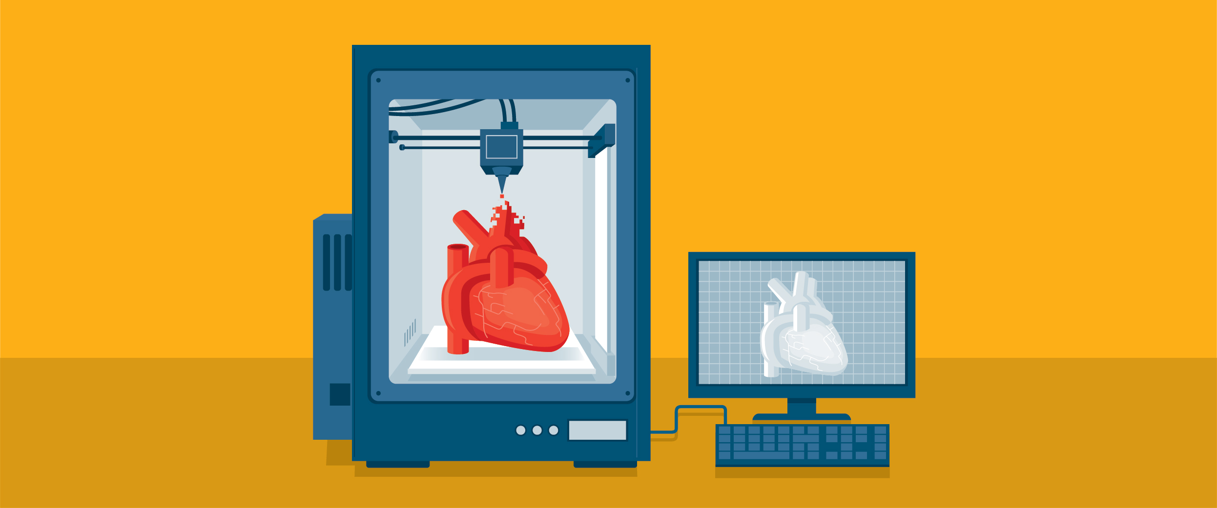
Biological 3D printing
Biological 3D printing, often referred to as 3D bio-printing, aims to generate 3D structures and organs using cells as well as inanimate materials. Some consider the pinnacle of 3D bio-printing as constructing entire functional organs from scratch that could effectively replace failing ones, someone no one has accomplished yet. However, MDs are already benefiting from 3D bio-printing, as it has been used to design intricate life-like models of organs, perfect for surgical planning and medical training (instead of relying on human cadavers). Furthermore, some have adapted the technology to printing realistic tumor-like models, which allows scientists to study tumor formation and development using in vitro 3D models rather than merely examining 2D plates filled with cancer cells. These models may also speed up the rate of drug efficacy and toxicity screening, especially since most pre-clinical screening currently depends on models that aren’t even human.1

So, how does one 3D print layers of cells?
It’s an exceedingly complex process, but the principle idea involves using “bio-ink,” which is composed of cells mixed with hydrogels (a type of gel with high water content), and producing a scaffold for the bio-ink to be deposited on. As it’s deposited, it’s crosslinked through light or heat activation.3
One of the main issues with bio-printing is maintaining the fidelity of the cells when they’re laid down so that they don’t lose their natural properties (e.g. ability to migrate) or die. Therefore, much work has gone into developing the materials and methods used for 3D bio-printing. Researchers have devised four distinct ways to bio-print:4
Inkjet-based bio-printing – Inkjet-based bio-printing can be achieved in one of two ways: either using a thermal element or a piezoelectric element, which generates electricity from mechanical stress. These elements eject droplets containing hydrogel-coated cells into a scaffold. Whereas thermal elements use heat to discharge the droplet, piezoelectric elements take advantage of acoustic waves for droplet expulsion. With inkjet bio-printing, you can print extremely detailed structures while keeping most of your cells viable.
Laser-based bio-printing – This technique relies on directed ultraviolet or visible light, which reacts with “photoinitiator” compounds that are mixed with cells. These photoinitiators are composed of chemicals that react with light, producing substances that can form polymers. While the absence of mechanical stress is a plus, potential UV-induced damage and the limited choice of photoinitiator molecules have made this method difficult to apply.
Laser-assisted bio-printing – This method uses a laser pulse to guide the passage of bio-ink through a donor-slide, which has a metal layer that becomes vaporized by the laser, to a receiver-slide, where the tissue is generated. Laser-assisted bio-printing yields a high degree of accuracy with regards to cell placement, but it’s expensive and can’t be scaled to large structures.
Extrusion-based bio-printing – Here, pressure is used to extrude bio-ink through a nozzle driven by mechanical or pneumatic pressure. This technique can use a high density of cells to print with, but with limited structure detail.
3D printing is relatively new, especially when it comes to biomedical research. Still, in its infancy, you can expect that new techniques will be developed (and current techniques will be constantly updated) to help make tissue engineering, disease modeling, and drug screening more efficient. One day, the technology might even be practical enough to replace failing organs with personalized structures built from scratch, reducing the waiting time for a healthy organ and perhaps removing the need for organ donors altogether.
LabTAG by GA International is a leading manufacturer of high-performance specialty labels and a supplier of identification solutions used in research and medical labs as well as healthcare institutions.

References:
- Paul GM, Rezaienia A, Wen P, Condoor S, Parkar N, King W, Korakianitis T. Medical Applications for 3D Printing: Recent Developments. Mo Med. 2018;115(1):75-81.
- Yan Q, Dong H, Su J, et al. A Review of 3D Printing Technology for Medical Applications. Engineering. 2018;4(5):729-742.
- Charbe N, McCarron PA, Tambuwala MM. Three-dimensional bio-printing: A new frontier in oncology research. World J Clin Oncol. 2017;8(1):21-36.
- Kačarević ŽP, Rider PM, Alkildani S, et al. An introduction to 3D bioprinting: Possibilities, challenges and future aspects. Materials (Basel). 2018;11(11):1-21.
