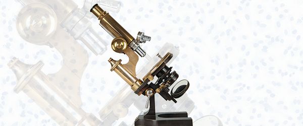
 Histology is one of the most varied fields of research, with a host of practical applications. Scientists have used the histological staining of tissues to understand how our bodies work, to discover novel therapeutic targets for disease, and to help diagnose patients suffering from illness. The term histology was coined in 1819 by Karl Mayer, who combined the two Greek words histos (tissues) and logos (study).1 However, the history of histology dates back even further with the advent of microscopy and the initial investigations into how tissues and organs work inside the body.
Histology in the 17th century
Histology, though not formally recognized as such at the time, began in the 1600s with Marcello Malpighi, who was born near Bologna, Italy in 1628. As a scientist, he experimented with insects, botany, and embryology. He was the first person to describe respiration in insects using the silkworm as a model, and he studied the development of chick embryos in more detail than anyone else had previously, making him a pioneer in the field of embryology. What he was most well-known for, however, was his characterization of pulmonary capillaries and alveoli. Malpighi visualized pulmonary capillaries in the frog—which he referred to as “the microscope of nature,” as it allowed him to see structures that were not visible in larger animals—using only a single magnifying lens. Though many of Malpighi’s discoveries were made using this lens, he was also aided by the invention of the compound microscope at the end of the 16th century, which allowed him to visualize chick embryo development. Notably, many structures within the human body are named after Malpighi, such as the Malpighian corpuscles, the Malpighian layer in the skin, and the Malpighian tubules in the excretory system of insects.2
The advent of modern histology
The idea that organs were composed of tissues wasn’t truly understood until the late 1700s when Marie-François Xavier Bichat introduced the term “tissue” to the medical lexicon and proposed that tissue within an organ may be impaired without the entire organ failing.3 He worked mostly by dissecting corpses, though he was steadfast in refusing to use a microscope, as he thought that the tool’s imperfections up to that point made it no better than chemical tests and the naked eye.3,4 However, around that time, J.J. Lister resolved to solve the inadequacies of the microscope by correcting its main issues: color fringes caused by chromatic aberrations and poor image quality caused by spherical aberrations and coma. His findings were only presented in 1830, though he had used his new microscope to perform histology long before that. Eventually, this led to the production of better microscopes throughout England and other countries, including Germany, which used them to fully establish the field of pathological medicine in the 1850s.5 Other advances made it easier for scientists to perform histological analysis, including permanent mounting in Canada balsam—perfected in 1851—and tissue fixation, which made it possible for scientists to interchangeably analyze samples without the specimen degrading.4
Development of staining techniques
Histological staining also dates to the 1700s. Carmine, a stain used by early botanists that has recently been replaced by the periodic acid-Schiff (PAS) technique, was developed by John Hill in the 1770s to study tissues in ammonia-based solutions. Hematoxylin, still one of the most widely used stains today, was first used by Wilhelm von Waldeyer in 1863 as a nuclear stain with a short staining time that was resistant to acidic solutions. Other commonly used stains developed in the 1800s include the Gram stain for bacteria (1875), Romanovsky-type stains (e.g. the Giemsa stain in 1891) for blood smears, the Zielh-Nielsen stain for tuberculosis (1883), and methylene blue (1891), which was initially used to treat malaria.6,7
Histology today
Performing histology today is a much different experience than it was at the turn of the 20th century. It was then that paraffin wax and formalin were first employed to embed and fix tissues, respectively, with the invention of a microtome capable of sectioning animal tissue occurring slightly earlier, in 1848. Important additions have also been made to histology procedures within the last several decades, such as the use of antibodies to stain tissue sections in a process called immunohistochemistry that was first described in the 1980s.7 Scientists have also turned to automation to process a larger number of samples with greater speed and precision and to perform multiple kinds of stains simultaneously.8 This rise in sample number has also led to the need for chemical resistant histology grade labels, to ensure samples are properly and securely identified. As the biomedical research industry evolves, so too does histological analysis, as it continually responds to new scientific and medical challenges for a growing (and aging) worldwide population.
LabTAG by GA International is a leading manufacturer of high-performance specialty labels and a supplier of identification solutions used in research and medical labs as well as healthcare institutions.
References:
- Hussein IH, Raad M. Once Upon a Microscopic Slide: The Story of Histology. J Cytol Histol. 2015;6(6):1-4. doi:10.4172/2157-7099.1000377
- West JB. Marcello Malpighi and the discovery of the pulmonary capillaries and alveoli. Am J Physiol Lung Cell Mol Physiol. 2013;304(6):L383-90. doi:10.1152/ajplung.00016.2013
- Shoja MM, Tubbs RS, Loukas M, Shokouhi G, Ardalan MR. Marie-François Xavier Bichat (1771-1802) and his contributions to the foundations of pathological anatomy and modern medicine. Ann Anat. 2008;190(5):413-420. doi:10.1016/j.aanat.2008.07.004
- Bracegirdle B. The history of histology: A brief survey of sources. Hist Sci. 1977:77-101. doi:10.1177/007327537701500201
- Bracegirdle B. J. J. lister and the establishment of histology. Med Hist. 1977;21(2):187-191. doi:10.1017/S0025727300037716
- Alturkistani HA, Tashkandi FM, Zuhair &, Mohammedsaleh M, Mohammedsaleh Z. Histological Stains: A Literature Review and Case Study. Glob J Health Sci. 2016;8(3):72-79. doi:10.5539/gjhs.v8n3p72
- Musumeci G. Past, present and future: overview on histology and histopathology. J Histol Histopathol. 2014:1-3. doi:10.7243/2055-091X-1-5
- Titford M. A Short History of Histopathology Technique. J Histotechnol. 2006;29(2):99-110. doi:10.1179/his.2006.29.2.99


