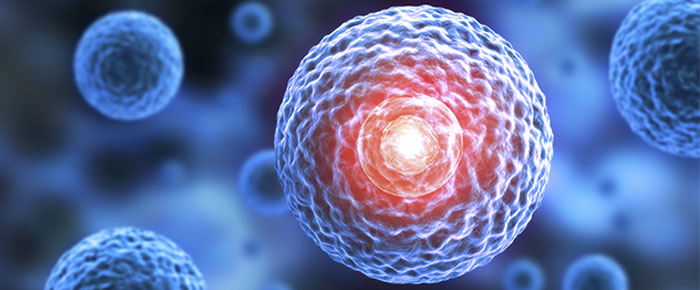
Cell culture is an invaluable tool in biology, drug discovery, cancer research, as well as in the study of stem cells. Currently, most labs culture in flasks or dishes using 2D methods. Though capable of providing insight into cellular signaling pathways and genomic regulation, among other things, 2D methods are limited by their in vitro nature. More recently, new 3D cell culture methods have been developed that have the potential to provide new insights and bridge the gap between 2D cell culture and animal models. These new 3D methods have improved upon cell culture’s utility in studying morphology, proliferation, stimuli responsiveness, differentiation, drug metabolism, and protein synthesis thanks to the ability to model a cell in vivo while being cultured in vitro2.
What is 3D Cell Culture?
3D cell culture artificially creates a three-dimensional environment in which biological cells are permitted to grow. The addition of a third dimension allows the environment to mimic complex interactions between cells and their extracellular matrix (ECM) not possible in traditional 2D cell culture. 3D cultures are usually grown in bioreactors, small capsules that allow the cells to grow into spheroids. When cultured in simple 2D Petri dishes or flasks, cells typically lose their original phenotypic and functional characteristics, modifying their responses and hampering their utility in drug discovery. In contrast, 3D cell cultures retain the characteristics of their in vivo counterparts, providing a more physiologically relevant research model.
Ideal 3D cell culture models promote cell proliferation and differentiation by enabling cell-to-cell and ECM interactions and providing oxygen, nutrient, and metabolic waste gradients. There are various models currently employed, including scaffold-based and scaffold-free techniques. These models have their own strengths and limitations, making them specific for only a select few applications. More generally, scaffold-based models more closely mimic cell-to-ECM interactions, while scaffold-free spheres are more amenable to cellular and physiological gradients.
Scaffold-Based Technologies
This model provides a physical support on which cells can aggregate, proliferate, and migrate. However, cells are typically embedded into the matrix, resulting in the chemical and physical properties of the scaffold influencing cell characteristics1. Scaffolds can be biological in origin or synthetic, which allows growth factors, hormones, or other molecules to be added in order to enhance cell proliferation or to promote a specific cell phenotype. Common scaffold-based models include hydrogel-based support, hydrophilic glass fiber, and organoids.
Anchorage-Independent Technologies
Scaffold-free 3D culture methods rely on cell self-aggregation in specialized culture plates that promote spheroid formation1. Spheroids imitate the physiological characteristics of tissue cell-to-cell contact and may allow for more natural cell-matrix interactions. Spheroid size depends on initial cell number; if large enough, they can display oxygen and nutrient gradients similar to tissue1. Tumor spheroids made using patient cells can even retain their original genotype over time, facilitating drug development and screening. The primary limitation of this model is the need to optimize culture conditions to enhance reproducibility while preventing the spheroids from getting too large to hinder nutrient supply and induce necrosis. Common scaffold-free models include hanging drop microplates, magnetic levitation, and spheroid microplates with an ultra-low attachment coating.
Applications in Drug Discovery
In recent years, 3D cell culture systems have become more prominent in drug discovery as they produce results with greater predictive value for clinical outcomes than traditional 2D models. In addition, as 3D models utilize human cells, they are preferred to certain animal models, which are not always able to accurately reproduce human diseases or capture specific side effects. While 3D cell cultures allow for the production of specific cellular environments that promote a desired cell behavior, more work is still needed to precisely represent in vivo conditions and disease pathology. Furthermore, cancer drug discovery models combining 3D cell culture with primary patient-derived tumor cells may pave the way for preclinical screening of a personalized drug panel, improving potential outcomes and reducing unwanted side effects1.
The consistency of response to drugs is the primary reason 3D cell culture models are gaining popularity for drug discovery2. This discrepancy in responses, between 3D and 2D cultures, can be attributed to various factors. One is that as 2D cells cannot maintain a normal morphology and are more susceptible to the effects of drugs. This is compounded by the organization of surface receptors in 2D cell cultures. The spatial arrangement of these receptors likely affects the binding efficacy of the drugs, eliciting responses that are not relevant to in vivo conditions. In contrast with 2D cultures, 3D cultures can be found in varying stages of development, with continual proliferation, a factor that is required for certain medications to be effective. Lastly, 3D cultures have greater depth, which may affect pH levels and, by consequence, drug efficacy.
The Future of 3D Cultures
Though 3D cell culture offers an array of advantages, it still has several limitations it must overcome before it can be widely adopted. It is currently more expensive than traditional cell culture methods and take more time to set up. Additionally, 3D cell cultures have proven difficult to image using conventional wide-field and confocal fluorescent microscopy2. Fluorescence microscopy is particularly challenging, as 3D cell cultures require multiple images to form a z-stack, which increases the imaging time significantly, as well as the need for computer storage space. Another commonly used technique, flow cytometry, which is used to count and sort cells, faces limitations when employed in 3D cultures. To integrate 3D cultures with flow cytometry, spheroids must be broken up into a single-cell suspension, and the cells must be disposed of following the completion of the assay.
Despite these challenges, many laboratories still plan on switching to 3D cell culture. As more make the switch, methodologies are likely to be optimized that overcome current limitations. As such, the future of 3D cell culture remains bright, particularly in the fields of drug discovery and cancer treatment. As 3D tumor models become more advanced, immunotherapy should also improve, and new drugs will be discovered to better treat cancer, no matter the type2.
LabTAG by GA International is a leading manufacturer of high-performance specialty labels and a supplier of identification solutions used in research and medical labs as well as healthcare institutions.
References:
- Langhans SA. Three-Dimensional in Vitro Cell Culture Models in Drug Discovery and Drug Repositioning. Front Pharmacol. 2018;9:6.
- Jensen C, Teng Y. Is It Time to Start Transitioning From 2D to 3D Cell Culture? Front Mol Biosci. 2020;7:33.


