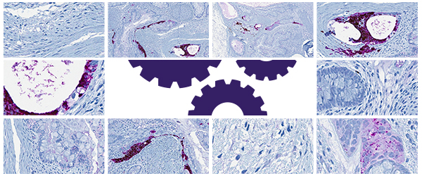
 Histology has evolved considerably since its beginnings in the 17th century, with advances in both specimen processing and analysis. Consequently, histology departments now face increasingly larger workloads. To adapt, they have integrated automated systems, which save time and allow histology professionals to work on other skill-based tasks while maintaining enough flexibility to process and stain according to the needs of the medical or research lab. Here, we’ll explore how automation has been integrated into histology to speed up the workflow of both medical technicians and researchers.
Automating tissue preparation
To produce slides for histochemical staining, tissues first need to be fixed, embedded in paraffin wax, and sections need to be cut. This entire process can now be automated by an array of different machines that perform each step of the process. Once the tissue is collected, it can be fixed using automated stations (e.g. the Milestone Medicine FixSTATION™) that control the temperature, time of fixation, and pH. Next, automated tissue processors can prepare specimens for embedding by performing all the necessary steps, including dehydrating the sample with increasing concentrations of ethanol and clearing it with a clearing agent (usually xylene, although some systems now come with xylene-free reagents, such as isopropanol). Some processors can monitor the concentrations of the reagents being used, notifying the user if there are any problems during sample processing. The samples are then treated with paraffin, though they must be still be embedded in molds, either manually or in a separate automated tissue embedder, prior to sectioning.
Automated tissue embedders come with reservoirs of paraffin and can properly orient the tissue sample, ensuring that it can be positioned in the proper three-dimensional plane for analysis, eliminating orientation errors and preventing the loss of tissue samples throughout the process. Once the blocks are solidified, they’re ready to be cut into sections using a microtome. Here, automation comes in the form of using a motorized microtome combined with a machine capable of performing automated section mounting. This tool allows you to take sections, relax the tissue in a water reservoir, then place the section onto a slide, reducing the likelihood of repetitive stress injuries due to repeated manual sectioning and mounting.
Slide staining and coverslipping
Slide staining is where automation necessitates the most flexibility, as it must be adapted for both basic histochemical as well as immunohistochemical analysis. There are two types of systems available: open and closed systems. Open staining systems provide the most flexibility and are often used for research purposes, where protocols may need to be customized depending on the tissue or biomarker. Closed systems provide less flexibility and are more suited for clinical labs, which prioritize reproducibility using established protocols.1 These systems may also come equipped with automated coverslippers that will dry your slides and immediately affix a coverslip to them.
Some of the features that can vary for automated slide stainers include slide and reagent management. For slide management, slides are loaded horizontally onto trays or racks and covered with reagent, which is spread around using either air or capillary action.1 However, the number of slides that can be processed at once varies, with some of the higher-end models capable of processing up to 330 slides per hour. Many machines, like the HistoCore SPECTRA ST from Leica, allow you to run high-throughput staining protocols for common stains like hematoxylin and eosin (H&E), while others, like the Ventana HE 600 system, allow you to stain individual slides, which is ideal for research labs with many users relying on differing staining protocols. With regards to reagent management, most systems use a matrix architecture, where both the slides and reagents are lined up in rows and columns and the robotic arm moves around dispensing reagents onto the slides. Reagent management can also be rotary, where the slides and reagents are organized around 2 rotating circular trays, and the rotation brings the slides into contact with the reagent-dispensing containers.1
Automated labelers
Specimens require identification and tracking throughout, from fixation to coverslipping. The best ways to do this are using either barcodes or radio-frequency identification (RFID), which can be scanned or read, as the samples are processed at each step. Many tissue processors, embedders, and slide stainers can scan barcodes (a few can scan RFID) and integrate the workflow into a management laboratory information system (LIMS). Some companies, like Leica, offer their own workflow management system called CEREBRO™, which can integrate with Leica and non-Leica–based systems.
When it comes to printing labels for histology, there are two options: automated printers for slides or cassettes and printing thermoplastic film labels. Using a thermal-transfer printer with xylene-resistant labels is the preferred choice as the printout from histology slide printers, which print directly on the glass of the slide, may not be as resistant and can smudge or fade during processing and staining. However, automated cassette printers are the superior option when it comes to the identification of tissue cassettes. Many different brands of all-purpose thermal-transfer printers are available, such as Citizen and Zebra. Several label printers, however, are tailored for histology automation, such as Leica’s Cognitive Cxi Label Printer, which scan the barcodes from tissue cassettes and print sets of labels for sample processing, embedding, and staining.
Within the last decade, automation has become a staple of histology. Depending on the needs of the lab, every step of the tissue preparation and staining process can be automated, reducing the time spent on processing and staining as well as providing more consistent results. Automation has even been implemented in histological analysis, with automated methods for stain normalization, tissue deformation correction, and image stitching.2,3 More innovation is sure to come in the near future, as the incorporation of artificial intelligence is set to overhaul how histology is performed, particularly with regards to data analysis, where massive amounts of data need to be compiled and sorted. Ultimately, the automation of histology will lead to faster and more accurate test results for the patients who depend on them and for the scientists working to find treatments.
LabTAG by GA International is a leading manufacturer of high-performance specialty labels and a supplier of identification solutions used in research and medical labs as well as healthcare institutions.
References:
- Prichard JW. Overview of automated immunohistochemistry. Arch Pathol Lab Med. 2014;138:1578-1582.
- Mccann T, Ozolek JA, Castro CA, et al. Automated Histology Analysis. IEEE Signal Process Mag. 2015;32(1):78-87.
- Van Eycke YR, Allard J, Salmon I, Debeir O, Decaestecker C. Image processing in digital pathology: An opportunity to solve inter-batch variability of immunohistochemical staining. Sci Rep. 2017;7:1-15.


