Phototoxicity can be a critical issue for those working with fluorescence, luminescence, and any other light-sensitive chemicals and cell cultures. Unfortunately, most labs are well-lit areas unsuitable for this kind of work. Here are several tips to help navigate such bright spaces and minimize the damage accrued by your light-sensitive samples and photosensitive reagents.
Use appropriate containers
Dark-colored, opaque vials and tubes are a must when storing light-sensitive chemicals. Ensure the containers block ultraviolet and visible light.
Keep solutions covered
For some assays, some tubes containing light-sensitive chemicals may need to be left open for a certain period. That doesn’t mean light needs to interact with the sample; instead, simply cover the samples with aluminum foil or place them within an enclosed box if long-term exposure is an issue.
Avoid working in well-lit areas when handling
Anyone who has used or currently uses buckets of chemicals to develop their Western blots will understand the necessity of working in poorly lit rooms. Similarly, if the chemical is so sensitive that any potential exposure, even for a second, can degrade it, it is worth investing in a dedicated space that can be fully darkened.
Employ light-protective reagents
Certain chemicals can protect against light within a solution, such as antioxidants, stabilizers, and inhibitors. One example is the commonly found and relatively cheap ascorbic acid. In a paper by Tomoki Harada from the University of Tokyo, they performed a screen for antioxidants that prevent photobleaching, concluding that ascorbic acid, at concentrations not cytotoxic to RPE1 cells, could optimally protect these cells from photobleaching during live-cell fluorescence imaging for mitosis.1 Other antioxidants that have the potential to reduce photobleaching include apigenin, chrysin, and beta-carotene.2 When employing light-protective reagents, always perform a quick test to ensure that the chemical does not unintendedly induce chemical degradation or cell death.
Track samples efficiently
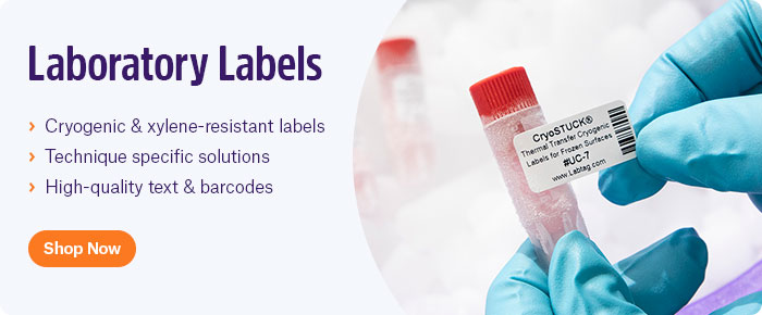
One of the most common ways photosensitive reagents become degraded is simply from repeated use, thus exposing them to light more so than fresh solutions. Without an efficient tracking method, it’s difficult to know how often an aliquot has been used and to what extent. To properly track these reagents, it’s recommended to use barcode and/or RFID labels in conjunction with a laboratory information management system (LIMS) or electronic notebook (ELN). Using barcode/RFID labels ensures that each sample, along with all relevant metadata like the number of times it has been used, volume, location, and concentration, are tracked in real-time. With a LIMS or ELN, this information can be readily accessed on a cloud-based system, so whoever requires the reagent can view the history of each individual tube or vial. Ultimately, this prevents lost time and money as it ensures that all reagents used in an assay, not just photosensitive ones, are validated prior to use.
When it comes to deciding between integrating barcodes or RFID, each lab must evaluate the pros and cons of each method. RFID does not require a direct line-of-sight and can scan multiple samples simultaneously, making it a much faster, though often costlier, option. While barcodes provide a more standardized method of sample tracking and usually require less troubleshooting, they can also be combined with RFID, as nearly any label can now be outfitted with an RFID inlay. These labels can be printed and encoded using a thermal-transfer RFID printer, providing maximum resistance against most laboratory environments.
LabTAG by GA International is a leading manufacturer of high-performance specialty labels and a supplier of identification solutions used in research and medical labs as well as healthcare institutions.
Reference:
- Harada T, et al. An antioxidant screen identifies ascorbic acid for prevention of light-induced mitotic prolongation in live cell imaging. Comm Biol. 2023;6:1107.
- Freitas JV, et al. Antioxidant role on the protection of melanocytes against visible light-induced photodamage. Free Rad Biol Med. 2019;131:399-407.


