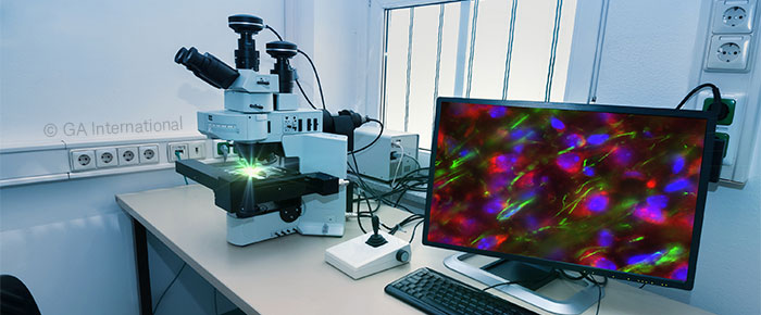
Histochemical analysis is an integral component of pathology laboratories, providing patients with diagnostic and prognostic information, which is critical for making treatment-related decisions. However, current standard-of-care technologies revolve around the use of 2D microscopy, which can provide only a slice of the true picture, especially regarding solid cancers. Though it is already popular in basic and translation science, 3D imaging appears to be on its way to the clinic as well, with the potential to revolutionize diagnostic healthcare.
2D vs 3D pathology
2D pathology typically involves the sectioning of patient biopsies following their fixation, dehydration, and embedding. The sections are mounted on slides to be stained and viewed via traditional 2D microscopy. Both human and AI approaches have been applied for slide analysis, but the idea remains largely intact: only a select section of the tissue sample is viewed at one time, providing limited information to the physician and patient.
Though 3D imaging has been around for many years, used throughout basic and translational research in the forms of confocal microscopy, multiphoton microscopy, and, more recently, light-sheet microscopy, its adoption in clinical diagnostics has been much slower. In 2016, several groups began to integrate these technologies for histopathologic purposes, with positive results regarding the ability of 3D imaging to stage cancers and assess patient prognosis.
Technological improvements in histopathology
Applying 3D imaging in the clinic would only be possible due to novel technologies that have been developed over the years:
Light sheet microscopy
Confocal and multiphoton microscopy has been a staple of scientific research labs for many years. However, because they generate images in a point-by-point manner, which necessitates scanning in three dimensions (X, Y, and Z planes), they tend to scan slowly and are complex to use. In contrast, light-sheet microscopy, also referred to as selective plane illumination microscopy (SPIM), has shown promise as a potential method with applicability for diagnostic labs. This form of microscopy generally utilizes transparent specimens, such as embryos and cleared tissues, to provide a fluorescent “scan” of one localized focal plane at a time. Fluorescence is only excited within a single detection plane at a time, thereby minimizing bleaching and photodamage.
In preliminary work shown at the ASCO 2023 Annual Meeting, one group from the Mayo Clinic in Rochester, Minnesota, integrated open-top light-sheet microscopy in combination with machine learning approaches to analyze a series of non-small cell lung cancer (NSCLC) specimens. In doing so, not only did they identify specific tumor cell types, but they were able to quantify additional features of the tumor, such as tertiary structures, stromal cells, and fibrosis.
Artificial intelligence
3D specimens generally provide a much higher data volume than 2D image slices. Not only is the data processing rate exponentially higher for 3D samples, but the intricacies inherent in properly detailing every component of a 3D structure require an ability to visualize and accurately quantify structures and provide an accurate diagnosis. Even in 2D pathology, it is recognized that machine learning can play a role in providing this type of support for clinicians.
One factor that needs to be addressed prior to the implementation of AI in 3D histopathology is the development of neural networks trained to use 3D data. Those currently being developed for 2D are not equipped to handle the complexity of 3D samples, nor are they trained to use them. Training sets will also be needed, with annotations provided by expert pathologists for the network to learn from. While this can be achieved within a long enough time frame, the primary challenge for AI in 3D pathology will be validating AI from a quality control standpoint.
Ultimately, if 3D imaging ever reaches the clinic, it is imperative that integration with other standard-of-care techniques occurs seamlessly, as 3D imaging is not likely to replace but rather supplement current diagnostics. However, once integrated, 3D imaging has the chance to revolutionize clinical decision making, providing a higher degree of certainty and understanding of a variety of diseases, particularly solid cancers.
LabTAG by GA International is a leading manufacturer of high-performance specialty labels and a supplier of identification solutions used in research and medical labs as well as healthcare institutions.
Reference:
- Liu JTC, et al. Harnessing non-destructive 3D pathology. Nat Biomed Eng. 2021;5(3):203-218.
- Stoltzfus CR, et al. Quantitative 3D assessment of the lung cancer microenvironment using multi-resolution open-top light-sheet microscopy. J Clin Oncol. 2023;41(16 Suppl):2616.