Specimen Collection
Specimens are traditionally collected from tissue samples removed from a living body, otherwise known as biopsies. The samples are often immediately placed in a temporary storage buffer until they reach the lab. At this step, proper labeling of the samples is of the utmost importance. Any sample ambiguity could result in a loss of time for the patient, the need to undergo additional surgery, as well as additional costs necessary to re-process the samples.
Biopsy specimens can be stored in a variety of containers, including small-diameter microtubes and vials, test tubes, plates, and bottles. Depending on the size and material of the collection container, a different type of label may be required. Note that many laboratories will not accept specimens whose identity is not affixed to the sample container.
Chemical Fixation
Once the cells are isolated, they must be fixed chemically, which is often performed with preservatives like formalin and glutaraldehyde. This freezes the tissues “in time” while also disabling enzymes that would normally break down the cells. As such, the tissue is protected from degradation while producing preserved cells that look very similar to their natural state.
At this step, any labels used to identify your samples might interact with a variety of different chemicals and solvents, such as ethanol, methanol, formalin, toluene, and more. Therefore, chemical-resistant labels are the ideal choice, with histology labels that can withstand formaldehyde and formalin solutions being particularly useful at this stage of the procedure.
Sample Dehydration & Clearing
Once fixed, cells must go through a dehydration series, which involves slowly replacing the water from the fixation buffer with increasing concentrations of alcohol. Following this, the ethanol is in turn replaced with xylene, a commonly used hydrophobic clearing agent, which will help once the wax embedding of the tissue begins.
Here, labels with an even greater chemical resistance are required as they are likely to come in contact with harsher solvents. Moreover, once sample dehydration is complete, the samples may require long-term freezer storage until they are ready to be processed. As such, labels that can withstand low temperatures and humid, damp environments are also recommended.
Paraffin Embedding
Once samples have been dehydrated and cleared, they are ready to be embedded in the embedding material (wax, agar, and resin). During this process, xylene is slowly replaced with increasing concentrations of the embedding material until no xylene remains, rendering the samples stable for several years. The most commonly used embedding material is paraffin wax due to its ease of use and its low melting point, which reduces tissue hardening. In electron microscopy, a harder matrix is necessary in order to cut very thin sections, requiring the use of resin instead.
Labeling of samples embedded in paraffin wax can be done directly on the wax block or the tissue cassette, with a chemical-resistant printout that will not fade or smear, using labels with an extra-permanent adhesive that allows the labels to adhere to hard to stick surfaces. For labeling tissue cassettes, a cassette printer is the preferable and most reliable option.
There are also labels designed specifically for the identification of resin-embedded samples. These labels are small enough to fit most resin capsules and molds, with strong resistance to chemicals and extreme heat. The labels can be used to securely label your samples with alphanumeric text or serialized numbers, as well as 1D or 2D barcodes.
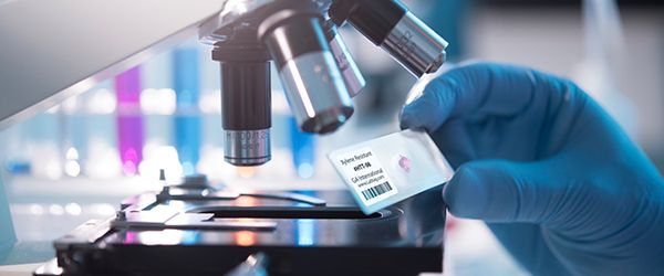
To stain samples, paraffin embedded samples must be thawed, and the blocks need to be thinly sectioned using a sharp blade, making thin cross sections in which to view the cells. Thin paraffin “ribbons” are affixed to microscope slides and extra wax is washed away by submerging the slide in xylene, which causes the wax to dissolve, leaving behind only the thin layer of tissue. Chemical stains, such as haematoxylin and eosin, are then applied to labeled histology slides in order to improve the contrast of the cell layers, making it possible to determine whether the patient has healthy or diseased tissues. Here, the use of labels designed to fit microscope slides, with a strong resistance to both direct contact and immersion in harsh chemicals and stains is essential. Slide printers are available as well, though durable stain-resistant labels still remain the more reliable choice.
Overall, the use of harsh chemicals, solvents, and stains in various histological processes requires that durable, chemical-resistant labels are used for proper and secure identification of samples. Luckily, LabTAG has developed a range of products to address this very need.
LabTAG by GA International is a leading manufacturer of high-performance specialty labels and a supplier of identification solutions used in research and medical labs as well as healthcare institutions.