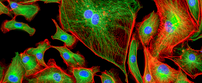
Starting with the invention of the microscope by a father and son team, scientists have been fascinated by the emerging new world of microorganisms. The advent of microscopy led researchers to want to record their findings, using simple hand-drawn images at first. This desire ultimately led to advancements in cinematography and the creation of short films showing the movement of bacteria. Today, live-cell imaging techniques have progressed much further and allow detailed videos of cells and their substructures to be recorded and studied.
Microcinematography
At the beginning of the 20th century, scientists started looking at the emerging film and cinema industry for ideas on how to record live cells. However, the French biologist Jean Comandon made the first significant scientific contributions around 1910 and is often cited as a pioneer of microcinematography. A trained microbiologist specializing in syphilis research, Comandon used an enormous cinema camera bolted to an ultramicroscope to record the bacteria’s movement that causes syphilis – Treponema pallidium1. He demonstrated that the movement of the disease-causing bacteria is uniquely different from the non-disease-causing form. He would go on to produce many other biological and medical films of cells, believing that recording cellular movement on film could help teach physicians to better understand and even diagnose certain diseases.
Interestingly, these early films of cells were shown in Paris theaters, running between newsreels and entertainment news. These movies also served to teach people about cells and molecules; with people excited, they could see something new they had never seen before.
The first films of bacteria and live cells were recorded in real-time; however, the flexible nature of film was soon used to enhance these faithful reproductions. Soon, scientists turned to time-lapse microcinematography. This form of microcinematography involved taking images at evenly spaced intervals and then playing them at a higher speed, thereby accelerating the perceived movements. This benefited from making previously imperceptibly slow changes visible to scientists, opening a new realm of biological phenomena to experimentation. Thus, live-cell microcinematography served as the precursor to modern-day live-cell imaging, driving forward scientists’ urge to see more, which has brought about advances in microscopy and video capture technologies.
Phase-contrast microscopy
The next giant leap in cellular imaging came during the early 1930s, with the phase-contrast microscope’s invention by Frits Zernike. A theoretical physicist by trade, his work with optics demonstrated how research in a highly specialized field can yield innovative developments in seemingly unrelated disciplines. With the help of Zeiss Optical Works in Jena, Germany, he designed the first phase-contrast microscope. However, it wasn’t long before most microscope manufacturers also started producing their own microscopes with this enhanced specimen illumination mode. In fact, the phase-contrast microscope proved to be such an advancement in microscopy that Frits Zernike was awarded the Nobel Prize in Physics in 1953.
By the 1930s, many staining techniques had been developed, though they all depended on the specimen being dead, limiting their usefulness. This new microscope allowed cellular details to be observed for the first time without using lethal stains. Light waves can cause the brightness and phase of the medium (e.g., cell) to change and give rise to colors, depending on the wavelength used. Traditional photographic equipment and the human eye are sensitive to changes in brightness, but phase changes cannot be perceived without special equipment. Phase-contrast microscopy allowed cellular structures that had been up to that point invisible by bright-field microscopes without the use of staining to become observable. This development made it possible for biologists to study living cells and how they proliferate through cell division.
The first recording of live cells using the phase-contrast microscope was done by Kurt Michel to present the meiotic cell division stages in living cells. He even began documenting the mitotic process involved in cell division by way of time-lapse cinematography in 1941. Modern phase contrast objectives can be couple with contrast-enhancing techniques to further improve cellular imaging. They can also vary absorption levels of the surrounding illumination to produce a broad spectrum of specimen contrast and background intensity.
Digital camera & modern imaging techniques
The next revolution in live-cell imaging came with the invention of the self-contained digital camera. Invented by Steve Sasson at Kodak in 1975, it offered excellent image quality, ease of use, and greater flexibility for image storage and manipulation.
The digital camera led to the rise of a number of new imaging techniques in the late 1980s that transformed our understanding of cell processes. This includes techniques such as widefield microscopy (WFM), confocal laser microscopy, and fluorescent imaging techniques (FRET, TIRF, FRAP, & FLIP). These new imaging techniques have allowed scientists to analyze the protein partners’ movement and interactions inside individual living cells.
Widefield microscopy (WFM)
WFM allows the entire specimen on the microscope stage to be illuminated2. This is the simplest and least expensive way to image live cells, ideally suited for monolayers of cells or thin slices. It commonly uses mercury arc-lamps and xenon arc-lamps as light sources, though light-emitting diodes (LEDs) have started being used instead recently. Post-processing procedures, such as deconvolution, can provide confocal quality images for thin specimens at a fraction of the cost.
Confocal laser scanning microscopy (CLSM)
The workhorse of every imaging lab, CLSM, uses laser units of varying wavelengths to image a small area of your sample, building the picture pixel-by-pixel by collecting the emitted photons2. This method’s primary advantage is that user-defined regions of interest can be selected for investigation, eliminating the need to scan the entire specimen. Moreover, CLSM can obtain optical sections through a sample, excluding much of the background fluorescence that may be out-of-focus.
Fluorescence imaging
There are multiple different fluorescence imaging techniques currently being used. Total internal reflection fluorescence microscopy (TIRF), also called evanescent wave microscopy, exploits the unique properties of an induced evanescent wave or field as a means to selectively excite cellular fluorophores immediately adjacent to the cell surface3. Furthermore, it can also reduce the interference of underlying regions within the cell or cellular structure. With many key events occurring in close proximity to membrane surfaces, this technique can significantly increase the number of quality images and information obtained.
Fluorescence localization after photobleaching (FLAP) enables tracking of molecules within living cells by utilizing localized photo-labeling3. The tracked molecule is given two fluorophores, a control and a target fluorophore that is rapidly photobleached at the chosen location. This allows the distribution of molecules to be located by simple image differencing and can characterize the mobility of cellular molecules.
The value of live-cell imaging innovation throughout history is magnified by the global Covid-19 pandemic and the inherent need for tools that can help us better understand and treat disease.
LabTAG by GA International is a leading manufacturer of high-performance specialty labels and a supplier of identification solutions used in research and medical labs as well as healthcare institutions.
References:
- Lorusso L, Lefebvre T, de Pastre B. Jean Comandon Neuroscientist. J Hist Neurosci. 2016;25(1):72-83.
- Cole R. Live-cell imaging. Cell Adh Migr. 2014;8(5):452-9.
- Dunn, G. A. et al. Fluorescence localization after photo bleaching (FLAP): a new method for studying protein dynamics in living cells. Journal of Microscopy 205, 109-112, 2002.