


Flow cytometry and fluorescence-activated cell sorting (FACS) represent essential techniques among immunologists and many other biologists. With the ability to quickly analyze and sort fluorescently-tagged cells, it is a mainstay for researchers and medical scientists. Here are several tips to help prepare and store flow cytometry/FACS samples that will help mitigate errors and increase consistency during the assay and before it has even begun.
Collect and store samples appropriately
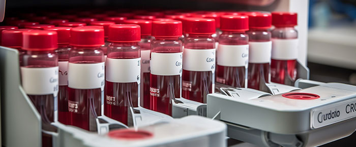
Collection and storage protocols will differ based on the types of cells being assayed, but the principle remains the same: use the right SOP and stick with it. Blood specimens and tissue samples may require specialized buffers and storage in deep-freeze temperatures as low as -80°C, especially when taken from clinical studies with multiple different sites. If cryopreservation is needed, don’t forget to ensure that suitable tubes and cryo labels are employed. If samples are collected and assayed fresh, general purpose labels may be utilized. However, pay close attention to ensure any labels affixed to flow cytometry assay tubes do not interfere with the path of the flow cytometer’s lasers.
Use automation
Cell counting is necessary for every FACS protocol to ensure the correct number of cells is counted in each tube. For years, manual counting using a hemocytometer and a microscope was the only method available to count cells. Newer methods have been devised since, with machines that perform automated counting, providing a more consistent and accurate representation of the cell density of the sample.
However, automated counting is the only thing that can be automated for FACS. For labs that perform a high volume of FACS sample preparation, automated labelers, and tube fillers are available that help reduce the labor-intensive work of affixing individual labels to each container. They can also come equipped with tube feeders, liquid handlers, and cappers/de-cappers to also speed up the process of setting up and running large assays.


Block to prevent nonspecific binding
Antibodies are known to be “sticky” when incubated with most types of cells. Just like you would when running a Western blot, it’s necessary to block cells prior to incubation with antibodies and subsequent FACS analysis. Either serum of the same animal the antibodies were developed in or a dedicated blocking solution can be employed to prevent nonspecific antibody binding. This is especially crucial if assaying monocytes or dendritic cells, as they typically express Fc receptors, which can bind antibodies via the Fc region rather than their antigen-specific binding site.
Measure viability
Dead cells can interfere with readings, showing up as nonspecific staining. Nuclei staining with propidium iodide is an easy way to ensure that the assayed cells do not have compromised membranes; however, it can only be used for live cells, not in fixed samples. For fixed samples, amine-reactive permeability dyes are recommended. These interact with intracellular proteins and withstand exposure to harsh fixatives.
Include controls
Every experiment and assay requires controls, and FACs is no exception. Here, controls are critical to validate results and monitor for nonspecific staining. Several types of controls are needed for each run: unstained controls to measure background fluorescence, compensation controls stained with only a single fluorophore to account for spectral overlap, and fluorescence minus one (FMO) controls, where all but one fluorophore is added so that fluorescence spread can be measured. Antibody isotype controls can also be used for antibody-related protocols to account for potential background staining from the antibody itself.
Optimize fluorophores
FACS cannot be optimized without precise knowledge of the exact excitation and emission wavelengths of the fluorophores you plan to stain cells with. To select the right panel of fluorophores, it’s worth noting the abundance of the antigens you wish to detect; those expressed at low levels may need a relatively bright fluorophore, whereas those expressed at high levels require a dim fluorophore. It is also important to avoid spectral overlap as much as possible.
LabTAG by GA International is a leading manufacturer of high-performance specialty labels and a supplier of identification solutions used in research and medical labs as well as healthcare institutions.


