What makes a good scientific model organism?
Model organisms are useful for understanding how specific processes occur in nature. To do so, scientists require the ability to do several things:
- Grow the organism
- Allow it to reproduce
- Visualize changes directly and via biochemical analysis
- Genetically and/or pharmacologically manipulate it
All common animal models fit these four requirements, and most have additional resources, such as a mapped genome and databases full of transgenic lines and other handy genetic tools. Though these models are easy to manipulate, they aren’t necessarily the best suited for addressing issues involving regeneration and evolutionary dynamics. Even mice are poor preclinical models for pharmacological efficacy in humans.
An interesting article about nontraditional model organisms was published recently by Benajmin Matthews and Leslie Vosshall, who represent a group of scientists that study a nontraditional model organism, the mosquito. In it, they describe the framework required to consider an organism as a proper model, comprising ten steps:1
Step 1 – Choose an organism that addresses pertinent scientific questions. The authors make sure to state that the choice of question—and therefore model—is a personal one, driven by the need to address a serious health issue or by mere curiosity to better understand how nature works.
Step 2 – Rear the organism in the lab. A bit of prior knowledge about the organism is necessary to work with a new model in the lab. This can include proper environmental conditions for growth and reproduction, such as traditional food sources and mating habits.
Step 3 – Profile its genome and gene expression. Though established centers might refuse to sequence the genome of a nontraditional model, it’s now possible to do this in-house using next-generation sequencing, as many have already done.2 To facilitate genetic manipulation, it’s also important to annotate the genome where possible and to perform RNA-Seq, which can be used to flesh out the transcriptome of the species.
Step 4 – Figure out how to introduce foreign genetic material. To genetically manipulate the organism, there must be a viable and efficient method to introduce vectors that can modulate gene expression. It’s worth considering whether these genetic mutations are to be heritable or somatic and to control for potential stress caused by the technique(s).
Step 5 – Develop a method for creating transgenic lines. This is necessary to drive the expression of markers and other proteins. There are several ways of doing this, including transposon-mediated transgenesis, site-specific integrases, and CRISPR knock-ins. Prior to developing a transgenic line, it’s often necessary to identify the right regulatory elements, so that the gene of interest will be efficiently expressed in the organism.
Step 6 – Knock-down gene expression. Though RNA interference (RNAi) can be used to knock down gene expression in specific tissues or during developmental stages, it tends to be transient and can result in variable levels of gene expression. For more permanent and consistent elimination of gene expression, genome editing via CRISPR is a more reliable technique.
Step 7 – Implement protocols for precise mutagenesis. While understanding the effects of gene knock-ins and knock-outs may yield pertinent information, it’s often necessary to selectively mutate specific genes in order to better understand the structure and function of its protein product. Homology-directed repair can be used to create many different kinds of mutants, including loss-of-function, in-frame mutations using exogenous sequences, and defined point mutations.
Step 8 – Design binary effector systems. These systems require a strain expressing an exogenous transcriptional activator that doesn’t attach itself to the host genome along with another strain carrying the reporter gene. When crossed together, the gene can then be expressed in any part of the organism at pre-determined time points.
Step 9 – Generate stable lines that can be used to visualize and manipulate tissues and cells. Using the tools established in the previous steps, strains can be generated that selectively express fluorescent protein or that directly allow certain tissues, like neurons, to be visualized and manipulated.
Step 10 – Grow the research! Start answering the questions you asked yourself before deciding to work with the new model and begin asking new ones as you start to understand the processes you’re studying. Your data can be extrapolated to understand biological principles across a more diverse set of organisms or to discover new techniques that could be applicable to traditional model organisms (even humans).
New model organisms to keep an eye on
Aedes aegypti (the common mosquito)
The article published by Matthews et al, though applicable to a wide range of nontraditional model organisms, was primarily focused on mosquitos, and with good reason. Mosquitos are the leading cause of animal-related death worldwide, spreading diseases like malaria, dengue fever, and the West Nile virus. Scientists like Matthews and Vosshall are now using mosquitos as a model for neurobiology, investigating the biological mechanisms that drive them to seek out blood and allow them to accurately locate hosts to feed on.1
Diatoms
Diatoms are strange creatures. They’re unicellular eukaryotes that can perform photosynthesis, but what makes them truly special is that their cell wall is made of silicon dioxide, or glass. This unique cell wall is the diatom’s most distinctive feature, as the pattern of silicon dioxide varies from species to species, carefully laid out in intricate shapes and different sizes. While the chemical basis for cell wall production is known—diatoms form silicon dioxide nodules from the conversion of silicic acid via silaffin proteins—what governs the shape and size of the wall has not yet been elucidated.2 The complete genome has so far been sequenced for two different species of diatoms.
Stentor and Axolotls
Both organisms offer different glimpses into a process critical for human health: regeneration. Stentor, a unicellular protist, can grow to over one millimeter, with a complex, polarized cellular structure. Interestingly, if you cut a stentor cell in half, it’ll regrow into two separate individuals, and lost body parts can even be grafted back into the cell. This makes it unique in terms of studying regeneration on a single-cell level.2
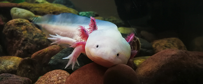
When it comes to tissue regeneration, one of the most interesting organisms to study is the axolotl. A type of salamander, the axolotl is a specialist in regenerating lost limbs. Because of how easy it is to rear the animal as well as its robust ability to reproduce (it lays 500 eggs at a time), the development of tools like CRISPR-mediated genome editing, which are required to manipulate the organism, are already available. As such, there’s been a recent burst of studies into how these animals can coordinate the regeneration of identical limbs so proficiently, taking advantage of mutant strains that express fluorescent proteins to help visualize the process.2
Tardigrades
There are few better multicellular organisms suited to studying resistance to extreme environments than tardigrades, also known as water bears. Tardigrades, with their eight legs and tiny size, can survive almost anywhere, from mosses and lichens, where they become desiccated, to cryogenic storage in liquid nitrogen and up to 4000 Gy of ionizing radiation. These organisms are being tested to identify what genes and molecules are responsible for making them so hard to kill, with several candidates being investigated that can protect both tardigrade and human DNA from damage.2
Choosing the right model organisms
The organisms listed above are only a handful of the many nontraditional organisms being used to study a variety of biological phenomena (for a more comprehensive list, see Russell et al 2020 in BMC Biology). What they all have in common is that they were chosen for specific reasons based on their biological characteristics and for their ability to be reared in the lab. However, while certain biological phenomena can be used to make a direct link to the question at hand (for instance, axolotl’s regenerate very well, so they make an ideal model to study regeneration), not all are capable of surviving laboratory conditions.
Bob Goldstein and Nicole King suggest taking a more pared-down approach to choosing the right animal. Instead of focusing on one species, pick a few that might help answer the same question, then narrow the search down based on practical considerations. In their experience with choanoflagellates, they tried growing every single species they could get their hands on until they had only two species that thrived in the lab. Using techniques developed for those two species, they adapted them to a wider range of choanoflagellates, slowly working their way towards understanding the evolutionary origins of multicellular development.3
LabTAG by GA International is a leading manufacturer of high-performance specialty labels and a supplier of identification solutions used in research and medical labs as well as healthcare institutions.
References:
- Matthews BJ, Vosshall LB. How to turn an organism into a model organism in 10 “easy” steps. J Exp Biol. 2020:1-10.
- Russell JJ, Theriot JA, Sood P, et al. Non-model model organisms. BMC Biol. 2017;15(1):1-31.
- Goldstein B, King N. The Future of Cell Biology: Emerging Model Organisms. Trends Cell Biol. 2016;26(11):818-824.