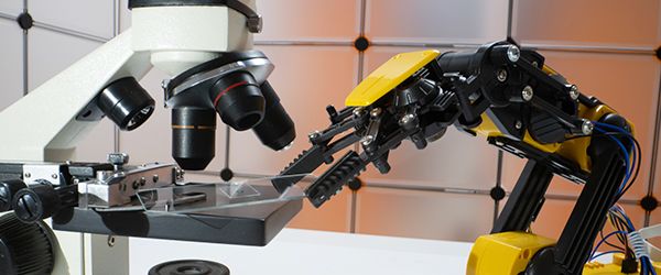
Application of AI in histological image analysis
One of the more recent technological breakthroughs in histology, whole slide imaging (WSIs), has made it possible to digitally process a slide after it’s stained using modern WSI scanners. However, such a large accumulation of data requires more time to analyze and study. AI is now being applied, to identify features in these images to help make diagnoses, to improve content-based image retrieval, and to find new clinically relevant associations within large pools of samples. These images can then be used for varied clinical applications that require either supervised or unsupervised learning. Supervised learning relies on providing the AI with training images so that it can match the appropriate characteristics of new images with those of previously annotated ones, which is critical when making a diagnosis. On the other hand, unsupervised learning can be used to interpret unlabeled images, extrapolating the data to find associations between clinical markers and disease. For example, Beck et al used AI to determine that stromal morphology correlates with the prognosis of patients with breast cancer.3
Limitations of AI
AI isn’t perfectly suited to take over the entire job of a pathologist, however. Though talk has centered around using AI as a sort of sidekick to the pathologist, there are still several hurdles towards making AI usable in the clinic.4 The main drawback to using any of the current iterations of AI in pathology and histology is the lack of publicly available training data.2 Annotated images are difficult to come by since only trained histologists or pathologists can accurately label them.3 Consider that within these databases, only a handful of diseases are represented, which would make it difficult for any program to efficiently learn what it needs to know to make an accurate diagnosis.3 There are several band-aid solutions to this problem (e.g. transfer learning, which involves applying the knowledge gained from one task to another), but the only true way to improve the performance of AI is to compile more training images.1
Another limitation is the cost of deployment; with pathology departments already under immense financial pressure, acquiring the required infrastructure to scan, store, and analyze gigapixel histopathological scans is a formidable challenge.2 Issues with no clear-cut solutions include trouble with multitasking appropriately as well as difficulties accounting for the subtler nature of the decisions the pathologist regularly makes, such as using cautious language or descriptive wording when making a diagnosis.2
AI may provide better care for patients
Assuming scientists can get AI running efficiently, it has the potential to significantly augment the value of patient data. It could also handle most of the pathologist’s and histologist’s benign, monotonous tasks like counting mitotic cells, leading to more efficient automation.2 Overall, AI could reduce the time for patients to receive a diagnosis by eliminating practical delays, such as triaging immunohistochemical tests and revisiting other cases, making it possible to select regions of interest and decide on follow-up tests immediately once the slide is processed.5
Many scientists are already beginning to refine their approaches to AI in pathology and histology. Some studies have already shown that AI can have an accuracy for diagnosing cases as high as 94%, though others are as low as 70%.1 Ultimately, it will be up to companies like PathAI and PAIGE to collaborate with physicians and develop a product―tailored for the needs of pathologists and histologists―that acquires approval from regulatory bodies, such as the US Food and Drug Administration (FDA), which would assure both patients and doctors that AI can be trusted to make appropriate clinical decisions.2
LabTAG by GA International is a leading manufacturer of high-performance specialty labels and a supplier of identification solutions used in research and medical labs as well as healthcare institutions.
References:
- Chang HY, Jung CK, Woo JI, Lee S, Cho J, Kim SW, Kwak TY. Artificial intelligence in pathology. J Pathol Transl Med. 2019;53:1-12.
- Tizhoosh H, Pantanowitz L. Artificial intelligence and digital pathology: Challenges and opportunities. J Pathol Inform. 2018;9(38):1-6.
- Komura D, Ishikawa S. Machine Learning Methods for Histopathological Image Analysis. Comput Struct Biotechnol J. 2018;16:34-42.
- Sharma G, Carter A. Artificial Intelligence and the Pathologist: Future Frenemies? Arch Pathol Lab Med. 2017;141:622-623.
- Djuric U, Zadeh G, Aldape K, Diamandis P. Precision histology: how deep learning is poised to revitalize histomorphology for personalized cancer care. npj Precis Oncol. 2017;1(1):1-5.